Table of Contents >> Show >> Hide
- What is emphysematous cholecystitis?
- How is it different from standard acute cholecystitis?
- Who is most at risk?
- What causes the gas?
- Symptoms: why it can fool people (and clinicians)
- How doctors diagnose emphysematous cholecystitis
- Treatment: what usually happens in the hospital
- Complications: why doctors take this so seriously
- Recovery and life after treatment
- Can you prevent emphysematous cholecystitis?
- Frequently asked questions
- Conclusion
- Experience Corner: what EC can feel like in real life (and what care teams often see)
- SEO Tags
Emphysematous cholecystitis is what happens when “regular” gallbladder inflammation decides it wants to be an
overachiever. It’s rare, aggressive, and has a flair for drama: gas forms inside the gallbladder wall or lumen,
usually from gas-producing bacteria. Translation: this isn’t the kind of belly pain you “sleep off” with herbal tea
and optimism.
This article breaks down what emphysematous cholecystitis is, why it’s dangerous, how it’s diagnosed (spoiler:
CT scans are the MVP), and what treatment typically looks likewithout turning the explanation into a medical
textbook that puts your browser to sleep.
Important: If you think you or someone else might have this condition, seek emergency care. This is not a DIY situation.
What is emphysematous cholecystitis?
Emphysematous cholecystitis (EC) is a severe form of acute cholecystitis (gallbladder inflammation) where gas is
present in the gallbladder wall, the gallbladder lumen, or nearby tissues. The gas usually comes from infection with
gas-forming organismsbacteria that basically “ferment” their way through tissue and generate air as a byproduct.
EC can progress quickly to complications like gangrene (tissue death), perforation (a hole in the gallbladder),
abscess formation, and sepsis. That’s why it’s treated as a surgical emergency more often than typical acute
cholecystitis.
How is it different from standard acute cholecystitis?
Standard acute cholecystitis is most commonly triggered by gallstones blocking the cystic duct, causing bile to back
up and irritate the gallbladder. In EC, the stakes are higher: reduced blood flow (ischemia) to the gallbladder wall
and infection by gas-forming bacteria are often part of the story. The result is a condition that can look similar at
firstbut behaves far more dangerously.
Key differences at a glance
- Speed: EC tends to worsen faster.
- Severity: Higher risk of gangrene, perforation, and sepsis.
- Imaging clue: Gas in or around the gallbladder is the “tell.”
- Outcomes: Reported mortality rates are higher than typical acute cholecystitis.
Who is most at risk?
EC can happen to anyone, but it’s seen more often in certain groups. The classic risk profile includes older adults,
people with diabetes, and individuals with vascular disease or compromised immunity. In many references, EC is also
described as more common in men than in women.
Commonly cited risk factors
- Diabetes mellitus (especially if long-standing or poorly controlled)
- Peripheral vascular disease or other conditions that reduce blood flow
- Immunosuppression (from illness or medications)
- Advanced age
- Severe acute illness or recent major surgery (sometimes linked to acalculous disease)
Why does diabetes show up so often? One theory: impaired circulation and immune response can make the gallbladder wall
more vulnerable to ischemia and infection, creating a welcome mat for gas-forming bacteria.
What causes the gas?
The gas typically comes from bacteria that can thrive in low-oxygen environments and generate gas as they metabolize.
Organisms frequently mentioned include Clostridium species (like Clostridium perfringens),
Escherichia coli, Klebsiella species, and sometimes anaerobes like Bacteroides.
A practical way to think about it: EC is often a two-hit problemreduced blood flow to the gallbladder wall plus an
infection that escalates quickly.
Symptoms: why it can fool people (and clinicians)
EC symptoms can overlap heavily with standard acute cholecystitis. That’s part of the danger: early on, it may not
announce itself with a neon sign that says “Hi, I’m the scary version.”
Common symptoms
- Right upper abdominal pain (may radiate to the right shoulder or back)
- Fever and chills
- Nausea and vomiting
- Abdominal tenderness (especially in the right upper quadrant)
- Sometimes jaundice (yellowing of skin/eyes), especially if bile ducts are involved
Some peopleespecially older adults or those with diabetesmay have less “classic” pain or a blunted fever response,
even while the condition is severe. Meanwhile, the infection can still be plotting its villain arc.
When to treat symptoms as an emergency
- Severe abdominal pain with fever
- Confusion, fainting, or extreme weakness
- Rapid heart rate, low blood pressure, or shortness of breath
- Worsening pain after initial improvement
How doctors diagnose emphysematous cholecystitis
Diagnosis usually combines clinical suspicion, lab tests, and imaging. Labs may show elevated white blood cell count
and inflammatory markers, and sometimes abnormal liver tests (especially if there’s bile duct involvement). But imaging
is where EC often gets unmasked.
Imaging tests used
- CT scan: Often considered the most sensitive test to detect gas in the gallbladder wall or lumen, and to evaluate complications.
- Ultrasound: Common first-line test for gallbladder issues; may show gallstones, wall thickening, and sometimes gas (though gas can make ultrasound harder).
- X-ray: Sometimes shows gas in the right upper abdomen, but it’s less sensitive than CT.
CT can help answer the big questions fast: Is there gas in the wall? Is there an abscess? Has perforation occurred?
Are nearby structures involved? In a condition where time matters, those details can steer life-saving decisions.
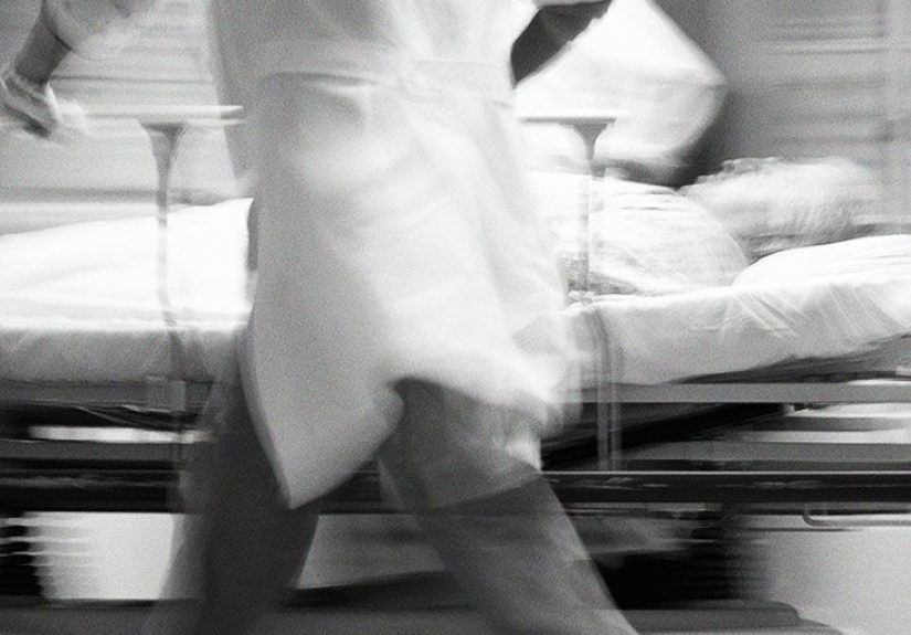
Treatment: what usually happens in the hospital
EC is generally treated urgently. Management depends on the patient’s stability, surgical risk, and the presence of
complications. Treatment typically includes resuscitation/supportive care, antibiotics, and definitive source control
(often surgery).
Step 1: Stabilize and support
- IV fluids and electrolyte management
- Pain control and anti-nausea medications
- Nothing by mouth (to rest the digestive tract)
- Monitoring for sepsis (vital signs, labs, urine output)
Step 2: Broad-spectrum antibiotics
Because EC can involve gas-forming organisms and potentially mixed aerobic/anaerobic bacteria, clinicians often start
broad-spectrum IV antibiotics promptly, then adjust based on cultures and clinical response. Antibiotics alone are
rarely the “full fix” in EC, but they’re crucial while definitive treatment is being arranged.
Step 3: Definitive treatment (source control)
In many cases, the preferred definitive treatment is urgent cholecystectomy (gallbladder removal),
often laparoscopic when feasible. However, when a patient is too unstable for surgery or is a very high surgical risk,
a percutaneous cholecystostomy (a drainage tube placed into the gallbladder through the skin) may be
used as a bridge or alternative, alongside antibiotics.
A realistic example timeline
Imagine a 68-year-old man with diabetes arrives with severe right upper quadrant pain and fever. Labs show elevated
white blood cells. Ultrasound suggests gallbladder inflammation, but the clinical picture seems “too sick” for
garden-variety cholecystitis. A CT scan reveals gas in the gallbladder wallclassic for EC. He receives IV fluids and
broad-spectrum antibiotics immediately. If stable enough, he goes for urgent cholecystectomy. If unstable, interventional
radiology places a cholecystostomy tube to drain infected bile and gas, while the team manages sepsis and reassesses
surgical timing.
Complications: why doctors take this so seriously
EC carries higher complication rates than standard acute cholecystitis. The gallbladder wall may become gangrenous,
and the risk of perforation is significantallowing infection to spill into the abdominal cavity.
Possible complications
- Gangrenous cholecystitis: tissue death due to severe inflammation and ischemia
- Perforation: a tear or hole in the gallbladder wall
- Pericholecystic abscess: localized pocket of infection near the gallbladder
- Sepsis and septic shock: systemic infection that can lead to organ failure
- Hepatic involvement: extension of infection into the liver in severe cases
Mortality rates reported in medical literature vary by study and patient population, but EC is consistently described as
having a higher mortality risk than uncomplicated acute cholecystitisone of several reasons rapid recognition and
treatment are emphasized.
Recovery and life after treatment
Recovery depends on how early EC is treated and whether complications occurred. After cholecystectomy, many people
gradually return to normal activities within weeks (sometimes sooner with minimally invasive surgery), while those
recovering from sepsis or perforation may need a longer hospital stay and more follow-up.
What “normal” can look like after gallbladder removal
- Most people can live without a gallbladder; bile still reaches the intestine, just less “stored.”
- Some people notice temporary digestive changes (especially with fatty meals).
- Diet adjustments are often short-term, but many patients do better long-term with moderate fat intake and smaller meals.
If a cholecystostomy tube was placed, follow-up is essential. Tube care, monitoring for infection, and planning for
eventual gallbladder removal (if appropriate) are common next steps.
Can you prevent emphysematous cholecystitis?
Because EC is rare and often associated with a sudden escalation of gallbladder disease, there’s no guaranteed “EC-proof”
plan. But you can reduce overall gallbladder risk and improve resilience.
Practical prevention strategies
- Manage diabetes well: better glucose control supports immune and vascular health.
- Don’t ignore gallbladder symptoms: recurring right upper abdominal pain after meals deserves evaluation.
- Address known gallstones when appropriate: follow clinician guidance on monitoring vs. treatment.
- General health maintenance: heart-healthy habits support vascular function, which matters for tissue perfusion.
Frequently asked questions
Is emphysematous cholecystitis contagious?
No. The infection is internal and related to gallbladder conditions and bacterial overgrowth, not person-to-person transmission.
Does EC always involve gallstones?
Not always. Many cases involve gallstones, but EC can also occur without stones (acalculous cholecystitis), particularly in very ill patients.
Why is a CT scan such a big deal here?
Because detecting gas in the gallbladder wall or lumenand spotting complications like perforationcan dramatically change urgency and treatment planning.
Can antibiotics alone treat EC?
Antibiotics are essential, but EC often requires drainage or surgery for definitive source control, especially when there’s gangrene, perforation, or sepsis risk.
Conclusion
Emphysematous cholecystitis is a rare but dangerous gallbladder infection characterized by gas in or around the gallbladder.
It can resemble routine acute cholecystitis early on, but it carries a higher risk of gangrene, perforation, and sepsis.
Rapid diagnosisoften with CT imagingand urgent treatment with IV antibiotics plus surgical removal or drainage are key.
If there’s one takeaway, it’s this: severe right upper abdominal pain with fever (especially in older adults or people with diabetes)
should be evaluated promptly. In EC, speed isn’t just helpfulit can be the difference between a difficult week and a dangerous outcome.
Experience Corner: what EC can feel like in real life (and what care teams often see)
Because emphysematous cholecystitis is uncommon, many people haven’t even heard of it until it’s suddenly in the room,
wearing a metaphorical trench coat and carrying a suspiciously large suitcase labeled “complications.” What follows are
common real-world patterns patients and clinicians often describegeneralized experiences, not individualized medical advice.
Patients often say the pain feels “different” from typical indigestion: deeper, sharper, and stubborn.
A classic story is pain in the right upper abdomen that doesn’t politely fade after a bathroom break or a nap. Some
people describe waves of nausea, a feverish “I feel poisoned” sensation, or the unsettling sense that something is
very wrong even if the pain isn’t the worst they’ve ever felt. In people with diabetes or in older adults, the pain
might be less dramatic than expectedyet the weakness, confusion, or rapid decline stands out. That mismatch can be
frightening: “I don’t feel that much painso why do I feel this sick?”
In emergency departments, the first challenge is often recognition. EC can masquerade as standard acute
cholecystitis. A clinician might notice that the patient’s vital signs look more concerning than the exam suggests:
faster heart rate, lower blood pressure, higher fever, or labs that hint at systemic infection. When the team orders
imaging, ultrasound may show inflammation but not explain the severityespecially if gas obscures the view. That’s
when a CT scan can change everything in minutes. Clinicians often describe the moment the CT report flags
intramural or intraluminal gas as an “all-hands” pivot: antibiotics are already running, and now surgery or drainage
planning accelerates.
Families frequently experience EC as a sudden sprint. One minute it’s “Maybe gallstones,” and the next
it’s “We need to treat this urgently.” That transition can be emotionally whiplash-inducing. It helps when teams explain
the logic clearly: gas-forming infection can damage the gallbladder wall quickly, and source control (removing or draining
the infected organ) prevents escalation to perforation or sepsis. When a patient is too unstable for surgery, a drainage
tube can look like a small procedurebut families often report feeling enormous relief when they hear, “We’ve decompressed
the infection and bought time.”
Recovery stories vary, but a common theme is “I didn’t realize how sick I was.” After treatmentespecially
once fever breaks and pain easesmany people are surprised by how quickly they feel clearer mentally and physically.
Others, particularly those who had sepsis or complications, describe a longer climb back: fatigue, reduced appetite,
and the need to rebuild strength gradually. Post-cholecystectomy, patients often share practical learning curves:
smaller meals feel better at first, greasy foods can be a gamble, and hydration becomes a quiet hero. Over time, most
find a new normal that feels, well, normal.
If you’re reading this because you’ve had ECor you’re caring for someone who hasknow that your reaction is valid.
It’s a scary diagnosis precisely because it moves fast. The upside is that modern imaging and emergency surgical/interventional
care are built for exactly this kind of problem: identify, stabilize, and control the source before it spirals.