Table of Contents >> Show >> Hide
- Table of Contents
- What a Mammogram Is (and Isn’t)
- Screening vs. Diagnostic Mammograms
- 2D vs. 3D Mammograms (Tomosynthesis)
- When to Start and How Often to Screen
- Benefits of Mammogram Screening
- Risks, Downsides, and Limitations
- Dense Breasts: Why They Matter
- What to Expect: Before, During, After
- Common Questions
- Real-Life Experiences: What People Often Say After Their Mammogram (and What They Wish They’d Known)
If you’ve ever heard someone describe a mammogram as “a quick pancake press,” they’re not entirely wrong.
But the real goal isn’t breakfastit’s catching breast cancer early, when treatment is usually simpler and outcomes are better.
This guide breaks down what a mammogram is, who should get screened (and when), what the benefits and risks really look like,
and how to make the whole experience less stressful and more “I’ve got this.”
Note: This article is for education, not personal medical advice. Your best screening plan depends on your age,
health history, family history, breast density, and overall riskso use this as a smart starting point for a conversation with your clinician.
What a Mammogram Is (and Isn’t)
A mammogram is a low-dose X-ray of the breast used to look for signs of breast cancer.
When it’s used in people who have no symptoms, it’s called a screening mammogram.
The big idea is simple: find cancer earlyoften before a lump can be felt.
What it isn’t: a mammogram is not a crystal ball. It won’t catch every cancer, and sometimes it flags something
that turns out to be harmless. That “not perfect” part is exactly why understanding benefits and risks matters.
Screening vs. Diagnostic Mammograms
Screening mammogram
This is the routine test for people at average risk with no breast symptoms.
It typically includes standard views of each breast, and the appointment is usually quick.
Diagnostic mammogram
This is the “let’s take a closer look” version. It may happen if:
- Your screening mammogram shows an area that needs extra images (“callback”).
- You have symptoms like a new lump, nipple changes, or persistent focal breast pain.
- Your clinician wants to evaluate a specific concern (including around implants or prior surgery sites).
A diagnostic visit may include extra mammogram angles (spot compression or magnification views) and often a same-day
ultrasound. Importantly, a callback does not automatically mean cancermost callbacks end up being benign.
2D vs. 3D Mammograms (Tomosynthesis)
You’ll often hear about 2D digital mammography and 3D mammography
(also called digital breast tomosynthesis).
2D mammography
Traditional digital mammography takes flat images of the breast. It’s widely available and has a long track record.
3D mammography (tomosynthesis)
Tomosynthesis takes multiple low-dose images from different angles and reconstructs them into thin “slices,”
which can make it easier to see through overlapping tissueespecially in dense breasts.
Many centers now offer 3D as standard, but availability and insurance coverage can vary.
Bottom line: 3D mammograms are commonly used and can reduce unnecessary callbacks in some settings,
but different medical groups interpret the evidence differently. If you have access to 3D, ask whether it’s appropriate for you,
what it costs, and how it may affect your chance of a callback.
When to Start and How Often to Screen
Screening guidelines can look confusing because different expert groups weigh benefits and harms differently.
The good news: most major U.S. organizations now agree that age 40 is a key starting point for
many people at average risk.
Quick comparison of common U.S. screening recommendations (average risk)
| Organization | Typical start age | Typical frequency | Typical end age | Notes |
|---|---|---|---|---|
| USPSTF | 40 | Every 2 years | Through 74 | Calls for more research for dense breasts and age 75+. |
| ACOG | 40 | Often every 1–2 years | Based on health/life expectancy | Emphasizes shared decision-making and individualized risk assessment. |
| American Cancer Society (ACS) | Option 40–44; start by 45 | Annual (45–54), then every 2 years (55+ optional annual) | As long as overall health is good | Encourages continuing if life expectancy is 10+ years. |
| American College of Radiology (ACR) | 40 | Often annual | Individualized | Also stresses risk assessment earlier in adulthood to identify high-risk patients. |
What “average risk” means
“Average risk” generally means you don’t have major risk factors like a known high-risk genetic mutation (such as BRCA),
a strong family history, or prior radiation to the chest at a young age.
Many people still develop breast cancer without a dramatic family history, which is why screening exists in the first place.
Who may need earlier or extra screening
If you’re at higher risk, your plan may include starting earlier, screening more often,
and/or adding tests like breast MRI.
Risk factors that commonly trigger a high-risk discussion include:
- Known inherited gene mutations (e.g., BRCA1/BRCA2) or strong family history.
- Prior chest radiation therapy at a young age (for example, for lymphoma).
- Very dense breasts plus other risk factors (dense tissue can both increase risk and make mammograms harder to read).
- Personal history of certain high-risk breast findings (your clinician will tell you if this applies).
Some high-risk guidelines recommend annual breast MRI plus mammography, often starting around age 30
(sometimes earlier depending on the risk factor). The key move is a formal risk assessmentnot guesswork.
Benefits of Mammogram Screening
The headline benefit is straightforward: screening mammography can find breast cancer earlier, which can:
- Reduce the risk of dying from breast cancer in screened populations.
- Increase the chance that treatment is less intensive (for example, smaller surgery or avoiding more aggressive therapy in some cases).
- Detect cancers before symptoms appear, when options are often broader.
Real-world example: the “nothing felt wrong” cancer
Many screen-detected cancers are found in people who feel completely normalno lump, no pain, no warning signs.
That’s the entire point of screening: you don’t wait for a problem to become obvious.
Another benefit people don’t talk about: clarity
While waiting for any medical result is nerve-wracking, a routine mammogram can also bring peace of mind.
For many people, the scariest part is the story they tell themselves before the testnot the test itself.
Risks, Downsides, and Limitations
Mammograms save lives, but they’re not free of tradeoffs. Understanding the downsides helps you make a decision that fits your values.
False positives and callbacks
A false positive means the mammogram suggests something might be wrong, but follow-up testing shows it’s not cancer.
This can lead to additional imaging (extra mammogram views, ultrasound) and sometimes a biopsy.
It can also cause anxietyyour brain doesn’t care that statistics are comforting when it’s 2 a.m. and you’re doom-scrolling.
Overdiagnosis (finding cancers that may never cause harm)
Screening can sometimes detect very slow-growing cancers (or pre-cancers) that might not have caused illness during a person’s lifetime.
The challenge is that we can’t always tell which findings will stay quiet and which will become dangerous,
so treatment often happens “just in case.” This is one reason recommendations differ on how often to screen.
False negatives (missed cancers)
Mammograms can miss cancersespecially in people with dense breast tissue.
If you notice a new breast change (like a persistent lump, skin dimpling, or nipple changes),
don’t let a “normal” screening result end the conversation. Screening is not a substitute for evaluating symptoms.
Radiation exposure
Mammograms use a small amount of ionizing radiation.
For most people, the benefit of early detection outweighs the radiation risk, but it’s still reasonable to keep screening appropriate
(not excessive) and to discuss the right schedule for your risk level.
Discomfort
The breast compression can feel uncomfortablesometimes briefly painfulbecause compression helps spread tissue for clearer images
and reduces the radiation needed. The good news: it’s typically quick, and technologists are used to adjusting positioning to help.
Dense Breasts: Why They Matter
“Dense breasts” means there’s more fibrous and glandular tissue compared with fatty tissue on a mammogram.
Dense tissue matters for two big reasons:
- It can make cancer harder to see on a mammogram (dense tissue and tumors can both appear white).
- It is associated with a higher risk of breast cancer compared with less dense tissue.
Starting in 2024, U.S. mammography facilities must provide standardized information about breast density in the patient notification,
and providers receive density information in the clinical report as well. If you’re told you have dense breasts, that’s not a diagnosis
it’s a data point. The next step is a risk conversation: do you need the same routine plan, a different interval, or supplemental imaging?
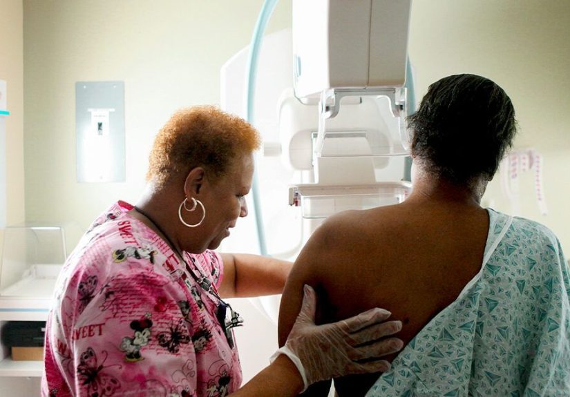
What to Expect: Before, During, After
Before your appointment: a preparation checklist
- Pick a less-tender time: If you menstruate, many clinicians suggest scheduling the week after your period when breasts may be less sensitive.
- Skip deodorant and powders: Avoid deodorant, antiperspirant, perfume, lotions, creams, and powders on your underarms or breasts the day of the test (they can show up on images).
- Wear a two-piece outfit: You’ll undress from the waist up.
- Bring prior images: If you’ve had mammograms beforeespecially at a different facilitybring or transfer the prior studies for comparison.
- Share key info: Tell the facility about implants, prior breast surgery/biopsies, pregnancy, or breastfeeding.
During the mammogram
A technologist positions each breast on the machine, then applies compression for a few seconds while the image is taken.
You may be asked to hold your breath briefly (yes, it’s a weird request for a breast test, but it helps reduce motion blur).
Most people are done in under 20 minutes, though times vary by facility and whether you’re having 2D, 3D, or diagnostic images.
After the mammogram: results and next steps
Results are typically reported using standardized language. You may hear about a BI-RADS category (a numbered scale that helps describe findings
and recommended next steps). For example:
- BI-RADS 0: More imaging needed (common after screening).
- BI-RADS 1–2: Negative or benign findings.
- BI-RADS 3: Probably benign; short-term follow-up may be recommended.
- BI-RADS 4–5: Suspicious; biopsy is often recommended to know for sure.
If you get called back, try to treat it like a request for “better photos,” not a diagnosis.
Follow-up testing is a normal part of screening, and most callbacks do not result in a cancer diagnosis.
Common Questions
Do mammograms hurt?
Some people feel pressure; some feel brief pain. Sensitivity varies.
Scheduling when breasts are less tender, communicating with the technologist, and staying relaxed can help.
If you’re very uncomfortable, speak upadjustments may be possible without sacrificing image quality.
What if I have breast implants?
You can still have mammograms. Let the facility know beforehand. Extra views may be needed to see as much tissue as possible.
What if I’m under 40?
Routine screening mammograms aren’t typically recommended for average-risk people under 40.
But if you have symptoms or significant risk factors, your clinician may recommend diagnostic imaging and/or earlier risk-based screening.
Do I need ultrasound or MRI too?
Not everyone. MRI is usually reserved for higher-risk screening because it can also increase false positives.
Ultrasound may be used as a follow-up tool or considered in certain risk profiles (including dense breasts),
but recommendations vary and evidence is still evolving for some groups.
Will insurance cover it?
Many health plans cover screening mammograms based on preventive-care requirements, but details differ by plan and by test type.
Ask about out-of-pocket costs for 3D mammography and any supplemental screening (MRI/ultrasound), which may have different coverage rules.
Real-Life Experiences: What People Often Say After Their Mammogram (and What They Wish They’d Known)
This section is based on common patient-reported experiences and what imaging teams frequently hearbecause the “how it feels”
part matters just as much as the “what it does” part.
1) “The anticipation was worse than the test.”
A lot of people walk into their first mammogram expecting a full-on ordeal, only to realize the actual imaging is short.
The most uncomfortable moments (compression and positioning) usually last seconds, not minutes.
What surprises many is how quickly it moves: check-in, quick instructions, a few images, and you’re back in your regular clothes
wondering why your brain scheduled a week of panic for a test that took less time than ordering coffee.
2) “I didn’t know deodorant mattered.”
This is the classic “I wish someone told me” detail. Many people show up wearing deodorant out of habit,
then feel annoyed (or embarrassed) when they’re asked to wipe it off. It’s not a judgment thingit’s a physics thing:
some products can leave tiny particles that show up on the images.
The practical fix is easy: skip it that morning, bring a travel deodorant for afterward, and consider it your small act of teamwork
with the radiologist.
3) “The callback freaked me out… and then it was nothing.”
Callbacks are emotionally loud. Even if you know intellectually that most callbacks aren’t cancer, your nervous system may not care.
People often describe a mental spiral: “What if… what if… what if…”and then the follow-up shows a cyst, a harmless overlap of tissue,
or a benign calcification pattern. If you’re called back, it can help to reframe it as a normal part of a high-sensitivity screening tool:
the system is designed to double-check anything even slightly unclear. Planning something grounding after your follow-up
(a walk, lunch with a friend, a calming playlist) can make the day feel less like a medical cliffhanger.
4) “Compression was uncomfortable, but the tech made a huge difference.”
People report that the technologist’s communication style changes everything. When the technologist explains what’s happening,
checks in during positioning, and gives clear “we’re almost done” cues, the experience feels manageable.
Many patients say the best move is simply speaking up: if something pinches, if your shoulder is straining,
or if you need a short pause, say so. Imaging teams do this all daythey’d rather adjust you than redo an image later.
5) “Seeing my report helped me feel in control.”
Some people prefer not to look at results until a clinician explains them; others feel calmer reading the summary
(especially the part that clarifies whether follow-up is needed).
Either way, a simple plan helps: decide in advance how you want results delivered, ask when to expect them,
and write down one or two questions for your cliniciansuch as how breast density affects your screening plan,
or whether your family history suggests a formal risk calculation.
6) “It became a routinelike a dental cleaning, but for my future self.”
Many people say the second mammogram is easier than the first because the fear of the unknown is gone.
Some even turn it into a low-stakes ritual: schedule it annually or every other year around the same time,
wear a comfy outfit, bring a podcast, and treat themselves afterward.
It’s not about pretending it’s fun; it’s about making it familiar. Familiar is calming.
If you take only one thing from these experiences, let it be this: you deserve a screening plan that fits your risk,
and you deserve an experience that respects your comfort. Ask questions. Bring support if you want it.
And remembergetting screened is not “looking for bad news.” It’s looking for information while you still have the most options.