
Table of Contents >> Show >> Hide
- What Is a CT Scan?
- What Is an MRI?
- CT Scan vs MRI: The Main Differences
- When Is a CT Scan Better Than an MRI?
- When Is an MRI Better Than a CT Scan?
- What to Expect During a CT Scan
- What to Expect During an MRI
- Safety Considerations: What Patients Should Tell the Imaging Team
- Cost and Availability
- Can You Need Both a CT Scan and an MRI?
- CT Scan vs MRI: Quick Comparison
- How Doctors Decide Which Test You Need
- Questions to Ask Before Your Scan
- Personal Experience-Style Insights: What the CT vs MRI Choice Feels Like
- Conclusion
If medical imaging had a personality contest, the CT scan would be the fast, no-nonsense friend who can find trouble in a hurry, while the MRI would be the detail-obsessed friend who notices the tiny wrinkle in your shirt from across the room. Both are powerful diagnostic imaging tools. Both help doctors see inside the body without making an incision. And both can sound mildly intimidating when your provider says, “We’re going to order some imaging.”
The good news? The differences between a CT scan and an MRI are easier to understand than they first appear. A CT scan, short for computed tomography, uses X-rays and computer processing to create detailed cross-sectional images. An MRI, short for magnetic resonance imaging, uses a strong magnetic field and radio waves to produce highly detailed images, especially of soft tissues such as the brain, spinal cord, muscles, ligaments, tendons, and internal organs.
In simple terms, CT scans are usually faster and excellent for emergencies, bones, bleeding, lungs, and certain abdominal problems. MRIs usually take longer but provide outstanding detail for soft tissue, nerves, joints, brain structures, and spinal conditions. Choosing between a CT scan vs MRI is not about which test is “better” overall. It is about which test is better for the medical question being asked.
What Is a CT Scan?
A CT scan is an imaging test that uses a series of X-ray images taken from different angles around the body. A computer then combines those images into detailed slices, almost like looking through a loaf of bread one piece at a time. Except, thankfully, nobody is calling your spleen sourdough.
CT scans are widely used because they are fast, accurate, and available in many hospitals and emergency departments. They can show bones, organs, blood vessels, and tissues with impressive clarity. A CT scan may be ordered to evaluate trauma, internal bleeding, fractures, lung disease, blood clots, kidney stones, tumors, infections, or abdominal pain.
Common Reasons Doctors Order CT Scans
A healthcare provider may recommend a CT scan when speed matters or when the body part being examined is best evaluated with X-ray-based imaging. For example, if someone arrives at an emergency room after a serious car crash, a CT scan may quickly help doctors look for internal injuries. If a person has sudden severe abdominal pain, a CT scan can help identify issues such as appendicitis, kidney stones, bowel obstruction, or internal bleeding.
CT is also useful for imaging the lungs. Because air-filled spaces and dense structures show up well on CT, doctors often use it to evaluate pneumonia complications, pulmonary nodules, chest trauma, and certain cancers. It is also used in cancer care to help locate tumors, stage disease, guide biopsies, and monitor treatment response.
What Is an MRI?
MRI stands for magnetic resonance imaging. Instead of using X-rays, an MRI uses a powerful magnet, radio waves, and a computer to create images of the inside of the body. The scanner encourages hydrogen atoms in the body to send signals, and those signals are translated into detailed images. That may sound like science fiction, but it is very real medicineand the machine is not judging you for Googling “MRI sounds like spaceship.”
MRI is especially strong at showing soft tissues. It can reveal details in the brain, spinal cord, nerves, muscles, ligaments, tendons, joints, blood vessels, and organs. Because it does not use ionizing radiation, MRI is often preferred when repeated imaging may be needed, although the final decision depends on the person’s condition, medical history, and the urgency of the situation.
Common Reasons Doctors Order MRIs
Doctors often use MRI to evaluate brain and spinal cord conditions, such as multiple sclerosis, tumors, stroke-related changes, spinal disc problems, nerve compression, and certain types of inflammation. MRI is also a favorite tool in orthopedics because it can show ligaments, cartilage, tendons, muscles, and joint structures better than many other imaging methods.
For example, if someone injures a knee while playing basketball and the doctor suspects a torn ACL, an MRI may be the preferred scan. A CT scan can show a fracture beautifully, but MRI can better show the ligament that decided to retire mid-game.
CT Scan vs MRI: The Main Differences
The biggest differences between a CT scan and an MRI come down to technology, speed, image detail, radiation exposure, comfort, cost, and the type of medical problem being investigated. Both tests can be lifesaving, but they are not interchangeable in every situation.
1. Technology: X-Rays vs Magnetic Fields
A CT scan uses X-rays. The scanner rotates around the body and captures multiple images, which a computer turns into cross-sectional views. Because X-rays are involved, CT scans expose the patient to ionizing radiation. Modern scanners and protocols are designed to use the lowest radiation dose needed to answer the medical question.
An MRI uses a strong magnetic field and radio waves. It does not use X-rays or ionizing radiation. This makes MRI attractive for certain patients and conditions, especially when detailed soft tissue imaging is needed. However, because MRI uses a powerful magnet, it requires careful screening for metal implants, medical devices, or metal fragments in the body.
2. Speed: CT Is Usually Faster
CT scans are usually much faster than MRIs. A CT scan may take only a few minutes, although preparation, contrast administration, and positioning can add time. This speed is one reason CT is so common in emergency medicine. When doctors need answers quickly, CT often gets called into action like a medical superhero with very practical shoes.
MRI exams typically take longer. Some MRI scans may last 20 to 60 minutes or more depending on the body part, the number of image sequences needed, and whether contrast is used. The patient must remain very still during the scan because movement can blur the images. For people who dislike enclosed spaces, that can feel challenging.
3. Image Strength: CT Loves Bones, MRI Loves Soft Tissue
CT scans are excellent for bones, fractures, lung imaging, bleeding, kidney stones, abdominal emergencies, and many trauma situations. They can produce clear images quickly and can also create 3D reconstructions, which may help surgeons plan procedures.
MRI is excellent for soft tissue contrast. It can show differences between normal and abnormal soft tissues in great detail. This makes it especially helpful for evaluating the brain, spinal cord, nerves, joints, tendons, ligaments, muscles, and certain organs. When doctors need to see subtle tissue differences, MRI often wins the staring contest.
4. Radiation: CT Uses It, MRI Does Not
One of the most important differences between CT and MRI is radiation exposure. CT scans use ionizing radiation because they are based on X-ray technology. For most medically necessary CT scans, the benefits outweigh the risks, especially in emergencies. Still, doctors consider radiation exposure carefully, particularly in children, pregnant patients, and people who may need repeated imaging.
MRI does not expose the patient to ionizing radiation. That does not mean MRI is risk-free for everyone, but its safety considerations are different. The main MRI concerns involve metal, implanted devices, heating effects, loud noise, claustrophobia, and contrast use in certain patients.
5. Contrast Material: Different Types, Different Questions
Both CT and MRI may use contrast material to make certain structures easier to see. CT contrast is often iodine-based and may be given through an IV, swallowed, or used in another way depending on the exam. It can help highlight blood vessels, organs, inflammation, tumors, or digestive tract structures.
MRI contrast is commonly gadolinium-based and is usually given through an IV. It can help doctors see inflammation, tumors, blood vessels, and abnormal tissue patterns. Contrast is not always necessary, but when it is used, it can add valuable detail.
Patients should tell their healthcare team about kidney disease, past contrast reactions, allergies, pregnancy, diabetes, medications, and any serious health conditions before either scan. Most people receive contrast safely, but careful screening helps reduce risk.
When Is a CT Scan Better Than an MRI?
A CT scan may be better when doctors need quick answers, especially in emergency situations. If someone has a suspected head injury, internal bleeding, major trauma, chest injury, or a possible fracture, CT often provides fast, useful information. CT is also commonly used for kidney stones because stones are usually visible on CT images.
CT may also be preferred for patients who cannot safely have an MRI because of certain implanted devices or unknown metal risks. Some pacemakers, cochlear implants, neurostimulators, metal fragments, or older surgical clips may create MRI safety concerns. Many modern devices are MRI-conditional, but the imaging team must verify safety before the scan.
Examples Where CT Often Helps
Imagine a patient who arrives in the emergency room with severe abdominal pain and fever. A CT scan can quickly help doctors look for appendicitis, abscess, bowel problems, kidney stones, or other urgent conditions. Another example is a patient who falls from a ladder and has severe hip pain. CT can show complex fractures that may be difficult to evaluate with regular X-rays alone.
CT is also helpful in certain cancer evaluations, especially for scanning the chest, abdomen, and pelvis. It can help doctors see whether a tumor has spread and whether treatment is working. In these cases, CT does not replace clinical judgment; it gives doctors a sharper map.
When Is an MRI Better Than a CT Scan?
MRI may be better when doctors need detailed images of soft tissue, nerves, the brain, the spine, or joints. It is often used for suspected ligament tears, spinal disc problems, brain tumors, multiple sclerosis, muscle injuries, pelvic conditions, and certain heart or liver evaluations.
MRI can also be helpful when doctors need to avoid radiation, especially if repeated scans are expected. However, MRI is not always the best choice in emergencies because it takes longer, requires strict safety screening, and may not be available as quickly as CT.
Examples Where MRI Often Helps
If a patient has persistent lower back pain with symptoms suggesting nerve compression, MRI can show spinal discs, nerves, and soft tissues in detail. If an athlete has shoulder pain and the doctor suspects a rotator cuff tear, MRI can help evaluate tendons and surrounding structures. If a neurologist needs to look for subtle changes in the brain or spinal cord, MRI is often the imaging tool of choice.
MRI can also provide detailed views of blood vessels through specialized techniques. In some cases, doctors may use MRI angiography to evaluate blood flow without using the same type of contrast used in CT angiography.
What to Expect During a CT Scan
During a CT scan, you usually lie on a narrow table that moves through a doughnut-shaped scanner. The machine may rotate around you while taking images. The scan itself is typically quiet and quick. You may be asked to hold your breath for a short time, especially during chest or abdominal imaging.
If contrast is used, you may feel a warm sensation, a metallic taste, or the urge to urinate, even though nothing dramatic is happening. This is common and usually passes quickly. The technologist watches from a nearby room and can communicate with you throughout the exam.
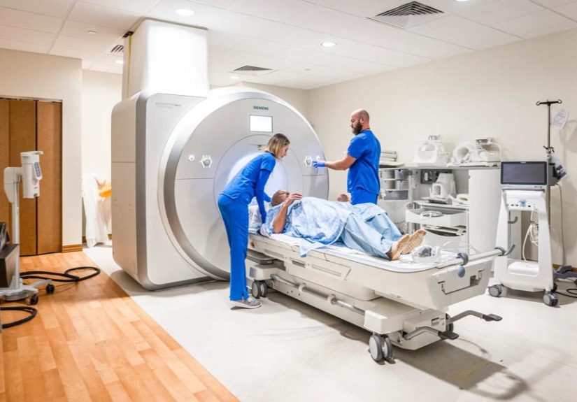
What to Expect During an MRI
During an MRI, you lie on a table that slides into a tube-shaped scanner. Some MRI machines are more open than others, but many still feel enclosed. The machine makes loud knocking, thumping, or buzzing noises, so earplugs or headphones are usually provided.
The most important job during an MRI is to stay still. Think of it as the world’s least exciting freeze dance. Moving can blur the images and may require repeating part of the exam. If you are claustrophobic, tell your provider before the appointment. Options may include calming techniques, music, open MRI, or medication when appropriate.
Safety Considerations: What Patients Should Tell the Imaging Team
Before a CT scan or MRI, the imaging team will ask questions to keep the exam safe. For CT, important details include pregnancy, kidney problems, diabetes, allergies, previous reactions to contrast material, and recent imaging tests. For MRI, patients must also report any implanted devices, metal fragments, surgical clips, artificial joints, pacemakers, cochlear implants, medication pumps, or other medical hardware.
Never assume a device is safe for MRI just because it sounds small. A tiny metal object can be a big deal around a powerful magnet. On the other hand, many implants are now MRI-conditional, which means MRI may be possible under specific conditions. The imaging team will check the details before proceeding.
Cost and Availability
Cost varies depending on location, insurance coverage, body part, contrast use, facility type, and whether the exam is done in a hospital or outpatient imaging center. In general, MRI is often more expensive than CT because the machines are complex, the exams take longer, and the image acquisition process is more detailed.
CT is usually more available in emergency settings. MRI machines may be less available after hours, and scheduling may take longer for non-urgent exams. Still, many hospitals and imaging centers offer both tests, and your healthcare provider will choose based on the clinical question rather than convenience alone.
Can You Need Both a CT Scan and an MRI?
Yes. Sometimes CT and MRI work as teammates. A CT scan may quickly identify an abnormality, and an MRI may later provide more detail. For example, a CT scan might detect a mass or injury, while an MRI helps define its relationship to nearby soft tissues. In cancer care, trauma, neurology, and orthopedic medicine, both tools may be used at different points in diagnosis and treatment planning.
This does not mean one scan “failed.” It means the body is complicated, and doctors sometimes need different tools to answer different questions. A hammer is great for a nail, but nobody wants a hammer used to tune a violin.
CT Scan vs MRI: Quick Comparison
| Feature | CT Scan | MRI |
|---|---|---|
| Technology | Uses X-rays and computer processing | Uses magnetic fields and radio waves |
| Radiation | Uses ionizing radiation | No ionizing radiation |
| Speed | Usually faster | Usually longer |
| Best for | Bones, bleeding, lungs, stones, trauma, emergencies | Brain, spine, nerves, joints, muscles, ligaments, soft tissue |
| Comfort | Shorter scan, more open machine design | Longer scan, louder, may feel enclosed |
| Contrast type | Often iodine-based | Often gadolinium-based |
| Metal concerns | Usually fewer metal-related restrictions | Requires careful screening for implants and metal |
How Doctors Decide Which Test You Need
Doctors choose between CT and MRI based on the suspected condition, the body part being examined, urgency, safety, medical history, and whether contrast is needed. They also consider whether the result will change treatment. A scan should answer a meaningful question, not simply create a fancy photo album of your insides.
For sudden trauma, suspected internal bleeding, or severe chest or abdominal symptoms, CT may be the first choice because it is fast and widely available. For a torn ligament, spinal cord problem, brain lesion, or subtle soft tissue abnormality, MRI may provide the better answer.
Questions to Ask Before Your Scan
Before having a CT scan or MRI, it is reasonable to ask your healthcare provider what the scan is looking for, whether contrast is needed, how the result may affect treatment, and whether there are alternatives. You can also ask how to prepare, whether you need to avoid food or drink, and when results are expected.
If you have kidney disease, implanted devices, pregnancy, claustrophobia, allergies, or a history of contrast reaction, mention it early. The more your care team knows, the better they can plan a safe and useful exam.
Personal Experience-Style Insights: What the CT vs MRI Choice Feels Like
For many patients, the difference between a CT scan and an MRI is not just technical. It is emotional. The doctor may understand the imaging decision perfectly, but the patient is thinking, “Will this hurt? Am I going to glow in the dark? Why does the MRI machine sound like a construction crew trapped inside a washing machine?” These are normal reactions. Medical imaging can feel mysterious when you are the person on the table.
One common experience with CT scans is surprise at how fast the exam can be. Patients often expect a long, complicated process, then discover that the actual scanning portion may be over quickly. The preparation can take longer than the scan itself, especially if IV contrast is needed. A person may lie down, slide through the scanner, hear a few instructions, hold their breath briefly, and then wonder, “That was it?” CT is not always simple, but compared with many medical tests, it can feel refreshingly efficient.
MRI, on the other hand, often feels more like an endurance event. The exam may require lying still for a longer period while the machine produces loud rhythmic sounds. Some patients describe it as annoying but manageable. Others find the enclosed space uncomfortable, especially if they already struggle with claustrophobia. This is why communication matters. Telling the imaging team about anxiety before the scan can make the experience much easier. Small adjustments, headphones, a mirror, a support person nearby when allowed, or medication prescribed in advance may help.
Another real-world difference is how each test fits into the diagnostic journey. A patient with sudden severe pain may receive a CT scan first because the doctor needs quick information. Later, if symptoms continue or more tissue detail is needed, an MRI may be ordered. This can worry patients who think, “Why do I need another scan? Did they miss something?” Usually, the answer is less dramatic. CT and MRI simply show different kinds of information. One may answer the urgent question, while the other answers the detailed follow-up question.
Patients also notice the difference in screening questions. Before a CT scan, the team may focus on contrast allergies, kidney function, pregnancy, and the reason for the exam. Before an MRI, the safety checklist can feel much longer because metal matters. You may be asked about pacemakers, implants, surgical clips, metal fragments, tattoos, hearing devices, medication pumps, and even past injuries involving metal. It may feel excessive, but the MRI magnet is powerful enough that caution is not optional. It is the kind of checklist you want the team to take seriously.
The best patient experience often comes from understanding the purpose of the test. CT is not “worse” because it uses radiation, and MRI is not “better” just because it sounds more advanced. A CT scan can be exactly the right test when speed matters or when bones, lungs, bleeding, or stones are the concern. An MRI can be exactly the right test when the issue involves soft tissue, nerves, the spine, or subtle changes in organs and joints. The smartest scan is the one that answers the medical question safely and clearly.
If you are scheduled for either test, the most practical advice is simple: ask what the scan is for, follow preparation instructions, disclose your medical history honestly, and speak up if you are anxious. Imaging teams do these exams every day, but for you, it may be your first time. A good technologist would rather answer one extra question than have you spend the entire scan silently rehearsing worst-case scenarios.
Conclusion
The differences between a CT scan and an MRI are rooted in how they work and what they show best. CT scans use X-rays, work quickly, and are especially useful for bones, lungs, trauma, bleeding, stones, and many emergency situations. MRIs use magnetic fields and radio waves, avoid ionizing radiation, and provide excellent detail for soft tissues, nerves, the brain, spine, joints, muscles, tendons, and ligaments.
Neither test is automatically superior. A CT scan can be the right choice when time matters. An MRI can be the right choice when detail matters. Sometimes doctors use both because each scan tells a different part of the story. The key is not to choose based on fear, guesswork, or whichever machine sounds more futuristic. The key is to match the imaging test to the medical question.
If your healthcare provider orders a CT scan or MRI, ask why it is recommended, what it may show, whether contrast is needed, and how the results may guide care. With the right information, medical imaging becomes less mysterious and much more useful. And honestly, anything that helps doctors understand what is happening inside the body without needing a crystal ball deserves a little respect.