
Table of Contents >> Show >> Hide
- Understanding the Basics: What Is Diabetic Macular Edema?
- The Main Cause: Long-Term High Blood Sugar
- Diabetic Retinopathy: The Road That Leads to DME
- VEGF and Inflammation: The Chemical Trouble-Makers
- High Blood Pressure: Extra Pressure on Fragile Vessels
- High Cholesterol and Abnormal Blood Lipids
- How Long Someone Has Had Diabetes Matters
- Kidney Disease and Fluid Balance
- Pregnancy and Rapid Changes in Diabetes Control
- Smoking: A Bad Deal for Retinal Blood Vessels
- Why DME Can Happen Even When Vision Seems Normal
- Diagnosis: How Eye Doctors Find the Cause
- Can Diabetic Macular Edema Be Prevented?
- Treatment Targets the Leak, Not Just the Symptom
- Real-Life Experiences: What Living With DME Can Feel Like
- Conclusion: The Cause Is Complex, but the Message Is Clear
- SEO Tags
Diabetic macular edema sounds like one of those medical terms designed to make normal people immediately want a snack and a nap. But underneath the fancy name is a very real eye condition that deserves attention, especially for anyone living with diabetes. In simple terms, diabetic macular edema, often shortened to DME, happens when damaged blood vessels in the retina leak fluid into the macula, the part of the eye responsible for sharp central vision.
The macula is the “high-definition screen” of the eye. It helps you read text messages, recognize faces, drive, cook, work on a computer, and spot whether your coffee mug is actually clean or just pretending. When fluid builds up there, vision can become blurry, wavy, faded, or distorted. The frustrating part is that DME may develop quietly at first, long before someone notices dramatic symptoms.
So, what causes diabetic macular edema? The short answer is long-term damage from diabetes-related changes in tiny retinal blood vessels. The better answer is more layered: high blood sugar, inflammation, blood pressure, cholesterol, diabetes duration, kidney disease, smoking, pregnancy, and delayed eye exams can all play a role. Let’s unpack the causes without turning this into a medical textbook wearing a lab coat.
Understanding the Basics: What Is Diabetic Macular Edema?
Diabetic macular edema is a complication of diabetic retinopathy, a diabetes-related eye disease that damages the small blood vessels in the retina. The retina is the light-sensitive tissue at the back of the eye. It works like a camera sensor, capturing visual information and sending it to the brain. When diabetes affects the retina, those tiny blood vessels may weaken, bulge, leak, close off, or trigger abnormal new vessel growth.
DME specifically occurs when fluid, proteins, and sometimes fatty deposits leak into or near the macula. This swelling thickens the retinal tissue and interferes with central vision. Peripheral vision may remain fairly normal, which can trick people into thinking nothing serious is happening. But if the macula is involved, everyday tasks can become harder very quickly.
The Main Cause: Long-Term High Blood Sugar
The biggest driver of diabetic macular edema is chronic high blood sugar. Glucose is necessary fuel for the body, but when blood sugar remains too high for too long, it can injure delicate blood vessels. The smallest vessels are often among the first to suffer, and the retina is packed with them.
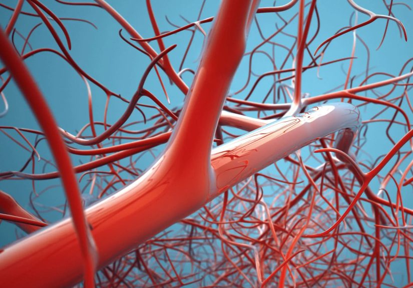
Over time, high glucose levels can damage the inner lining of retinal blood vessels. These vessels may become weak and leaky. Imagine an old garden hose with tiny cracks: it still carries water, but it also drips where it should not. In the retina, that “drip” is fluid leaking into retinal tissue. When the leak affects the macula, diabetic macular edema can develop.
High blood sugar also contributes to oxidative stress and inflammation. These processes can disrupt the blood-retinal barrier, which normally helps keep fluid and unwanted substances out of the retina. Once that barrier becomes less reliable, fluid can escape into the macula more easily.
Diabetic Retinopathy: The Road That Leads to DME
Diabetic macular edema usually does not appear out of nowhere. It is closely tied to diabetic retinopathy. In early diabetic retinopathy, known as nonproliferative diabetic retinopathy, tiny bulges called microaneurysms may form in weakened retinal vessels. These microaneurysms can leak blood, fluid, and lipids.
As diabetic retinopathy progresses, some vessels may close off, reducing oxygen delivery to parts of the retina. In response, the eye may release chemical signals that encourage the growth of new blood vessels. Unfortunately, these new vessels are fragile and poorly constructed. They are more like emergency scaffolding than sturdy architecture, and they can leak or bleed.
DME can occur at different stages of diabetic retinopathy. A person does not always need to have advanced retinopathy to develop macular edema. This is one reason regular dilated eye exams matter even when vision seems fine.
VEGF and Inflammation: The Chemical Trouble-Makers
One important factor behind diabetic macular edema is vascular endothelial growth factor, better known as VEGF. VEGF is a protein the body uses to help grow blood vessels. That sounds useful, and sometimes it is. But in diabetic eye disease, too much VEGF can make retinal blood vessels leakier and can encourage abnormal vessel growth.
Inflammatory chemicals also contribute to DME. Diabetes can create a chronic low-grade inflammatory state in the retina. This inflammation makes blood vessels more permeable, meaning fluid can pass through vessel walls more easily. Think of it as the retina’s security system getting too tired to check IDs at the door.
This is why many DME treatments target either VEGF or inflammation. Anti-VEGF injections help reduce leakage and swelling. Steroid treatments may be used in certain cases to calm inflammation. Treatment choice depends on the patient’s eye findings, medical history, response to prior therapy, and the eye specialist’s judgment.
High Blood Pressure: Extra Pressure on Fragile Vessels
High blood pressure is another major contributor to diabetic macular edema. When blood pressure runs high, it puts extra stress on blood vessel walls throughout the body, including the retina. If diabetes has already weakened those vessels, hypertension can make leakage more likely.
For people with diabetes, blood sugar control gets a lot of attention, and rightly so. But blood pressure deserves a seat at the same table. Poorly controlled hypertension can speed up diabetic retinopathy progression and worsen macular swelling. Managing blood pressure is not just about protecting the heart and kidneys; it is also about protecting the ability to read street signs and enjoy a good movie without squinting like a detective in a foggy alley.
High Cholesterol and Abnormal Blood Lipids
Cholesterol and triglycerides may also influence diabetic macular edema. When retinal vessels leak, fatty substances from the blood can settle in the retina as hard exudates. These yellowish deposits are signs that leakage has occurred. High lipid levels may increase the amount of lipid material available to leak into retinal tissue.
Not every person with DME has high cholesterol, and not every person with high cholesterol develops DME. However, abnormal blood lipids are often considered part of the bigger risk picture. Good diabetes care usually includes monitoring cholesterol, not only to lower cardiovascular risk but also to support overall vascular health.
How Long Someone Has Had Diabetes Matters
The longer a person has diabetes, the higher the chance of developing diabetic retinopathy and diabetic macular edema. This is true for both type 1 and type 2 diabetes. Duration matters because blood vessels experience years of exposure to glucose swings, inflammation, and metabolic stress.
Some people feel discouraged when they hear this, as if the calendar itself is the villain. But duration is only one part of risk. Many people who have lived with diabetes for years maintain strong vision through regular eye exams, careful glucose management, blood pressure control, and timely treatment. The goal is not perfection. The goal is consistent protection.
Kidney Disease and Fluid Balance
Diabetes-related kidney disease can be associated with diabetic eye disease. The kidneys and eyes both contain delicate small blood vessels, so damage in one area may reflect broader microvascular stress in the body. When kidney function is reduced, fluid balance, blood pressure, and inflammation may become harder to control.
Patients with diabetic kidney disease may therefore need especially careful monitoring for eye complications. This does not mean kidney disease automatically causes DME, but it can be part of the same vascular story. In diabetes, the body rarely files problems into neat little folders. The eyes, kidneys, nerves, heart, and blood vessels often share the same messy inbox.
Pregnancy and Rapid Changes in Diabetes Control
Pregnancy can increase the risk of diabetic retinopathy progression in some people with preexisting diabetes. Hormonal changes, blood volume changes, and shifts in blood sugar control may affect the retina. People with diabetes who are pregnant or planning pregnancy should talk with their medical team about eye exams before and during pregnancy.
Another important point: very rapid improvement in blood sugar control may temporarily worsen diabetic retinopathy in some patients, especially if retinopathy is already present. This does not mean improving blood sugar is bad. It means glucose goals should be managed thoughtfully with medical guidance, especially in people with known eye disease.
Smoking: A Bad Deal for Retinal Blood Vessels
Smoking damages blood vessels, reduces oxygen delivery, increases inflammation, and raises the risk of many diabetes complications. For the retina, this is not exactly a friendly care package. Tobacco use may worsen vascular injury and make it harder for the body to maintain healthy circulation.
Quitting smoking is one of the most powerful steps a person can take for overall health. It supports the heart, lungs, kidneys, nerves, and eyes. No one needs another lecture from the “health police,” but when it comes to diabetic macular edema, smoking is like adding glitter to a spill: it makes a bad situation harder to clean up.
Why DME Can Happen Even When Vision Seems Normal
One of the sneakiest things about diabetic macular edema is that early changes may not cause obvious symptoms. A person may still pass through daily life without noticing much difference. The brain is also excellent at filling in visual gaps. It is basically the world’s most confident photo editor.
Symptoms may appear gradually and can include blurry central vision, wavy lines, dull colors, difficulty reading, trouble seeing faces clearly, or dark spots near the center of vision. Sometimes one eye is affected more than the other, so the better eye compensates. This is why relying only on symptoms is risky.
Diagnosis: How Eye Doctors Find the Cause
An eye care professional can detect diabetic macular edema through a comprehensive dilated eye exam. During dilation, the doctor can examine the retina and look for signs of leakage, swelling, bleeding, abnormal vessels, or lipid deposits.
Optical coherence tomography, or OCT, is commonly used to measure retinal thickness and see fluid in the macula. It creates detailed cross-sectional images of the retina, almost like a tiny architectural scan of the eye. Fluorescein angiography may also be used. In this test, dye is injected into a vein, and special photos show where retinal vessels are leaking or blocked.
These tests help determine whether swelling is present, how severe it is, and what treatment may be appropriate. They also help track whether treatment is working over time.
Can Diabetic Macular Edema Be Prevented?
Not every case can be prevented, but risk can often be reduced. The strongest prevention strategy is good overall diabetes management. That includes keeping blood sugar within the target range recommended by a health care professional, controlling blood pressure, managing cholesterol, staying physically active when possible, eating a balanced diet, avoiding smoking, and attending regular eye exams.
Annual dilated eye exams are commonly recommended for many people with diabetes, though some need more frequent follow-up depending on their eye findings. People with type 2 diabetes should usually have an eye exam soon after diagnosis because diabetes may have been present for years before it was discovered. People with type 1 diabetes are generally advised to begin regular screening after several years of disease duration, based on medical guidance.
Treatment Targets the Leak, Not Just the Symptom
Treatment for diabetic macular edema aims to reduce swelling, stop leakage, and protect vision. The most common modern treatment is anti-VEGF medication injected into the eye. Yes, the phrase “eye injection” sounds like it belongs in a horror movie, but in real clinics, the eye is numbed, the process is quick, and many patients tolerate it far better than expected.
Other options may include corticosteroid injections or implants, laser therapy, and in selected advanced cases, surgery. Treatment is individualized. Some people improve quickly; others need repeated therapy over time. DME is often managed as a chronic condition, not a one-and-done repair job.
Real-Life Experiences: What Living With DME Can Feel Like
People often describe diabetic macular edema not as sudden darkness, but as a slow change in visual confidence. One day, reading a menu feels slightly harder. A phone screen needs to be brighter. Street signs look fuzzy until the car is closer. Straight lines on a spreadsheet may seem a little bent. At first, it is easy to blame fatigue, age, lighting, or the tiny font choices of modern civilization, which honestly should be investigated.
A common experience is the “good eye, bad eye” problem. Someone may cover one eye by accident while rubbing their face and suddenly realize that the other eye is much blurrier. Because both eyes work together, one eye can hide the weakness of the other for a surprisingly long time. This is why many people are shocked when an eye exam reveals swelling they did not know was there.
Another experience is emotional frustration. Vision is personal. It affects independence, work, hobbies, transportation, and confidence. A person who loves reading may feel betrayed when words blur. Someone who cooks may struggle to read labels. A grandparent may worry when faces do not look as crisp. These changes can feel scary, especially when the word “diabetes” has already taken up too much space in life.
Many patients also describe treatment anxiety. The idea of eye injections can sound impossible before the first appointment. But after the first few visits, many people say the anticipation was worse than the actual treatment. The numbing drops help, the procedure is usually brief, and the goal is meaningful: preserve vision. Some compare it to going to the dentist, except with fewer lectures about flossing.
Daily life with DME often becomes a partnership between medical care and practical habits. Patients may use larger fonts, better lighting, magnifiers, anti-glare screens, or voice features on phones. They may plan rides after appointments if vision is blurry from dilation. They may track blood sugar more carefully, take blood pressure seriously, or finally schedule that primary care follow-up they had been politely ignoring.
Support matters too. A family member who understands DME can help with transportation, appointment reminders, medication organization, or simply patience. Vision changes can make people feel embarrassed, but there is nothing embarrassing about protecting eyesight. The smartest move is early action. DME is not a character flaw, and it is not a punishment. It is a medical complication that can often be treated, monitored, and managed.
Conclusion: The Cause Is Complex, but the Message Is Clear
Diabetic macular edema is mainly caused by diabetes-related damage to tiny retinal blood vessels. High blood sugar weakens and inflames those vessels, making them leak fluid into the macula. High blood pressure, abnormal cholesterol, long diabetes duration, kidney disease, smoking, pregnancy, and delayed eye care can all add fuel to the fire.
The encouraging news is that DME is not invisible to modern medicine. Eye doctors can detect it with advanced imaging, and treatments can reduce swelling and protect vision. The earlier it is found, the better the chances of preserving sight. For anyone living with diabetes, regular eye exams are not optional fine print. They are one of the best tools for keeping the world sharp, colorful, and readable.
SEO Tags
Note: This content is for general educational purposes only and should not replace diagnosis, treatment, or personalized guidance from an ophthalmologist, retina specialist, optometrist, or diabetes care professional.